Monday, May 5, 2014

Battles Under the Sea

            A battle under the sea brings up iconic images; sharks and whales and other large animals fighting for their lives. However, this battle occurs deeper than what these animals could reach; roughly one mile below the ocean to be specific. In the deep darkness of the bathypelagic, a war is happening…a microbial war. It’s been recently discovered that viruses are attacking hydrothermal bacteria with one goal, their stores of sulfur.

A typical hydrothermal vent 
(Photo credit: NOAA)

            Bacteria and viruses have been enemies for as long as both have existed, but this is anew an interesting front in these battles. Deep-sea hydrothermal bacteria are still very unusual and not very well-known, and this is the first time that this sort of virus-bacteria relationship has been seen, since chemosynthetic bacteria are a rare lot.
            So, why do these bacteria target the sulfur in these bacteria? Well, the answer lies in the viruses’ desire to reproduce; turning the bacteria into a virus factory in a sense. What they do is they will force the bacteria to consume all of its sulfur reserves, and then use the resulting energy to create as many viruses as possible. The bacteria will inevitably die from this process, releasing all of the newly created viruses into the open to repeat the process.
            Interestingly enough, this sort of relationship has been seen before, namely in the shallow ocean water. There is a similar virus that target photosynthetic bacteria in order to reach the same end goal. So, it’s interesting to see the parallel between the two viruses, especially when it comes to their mechanism for reproducing.
            So, what are researchers doing about this? Well, currently Gregory Dick, a University of Michigan microbiologist and oceanographer, and his team are currently analyzing the DNA of the bacteria and viruses in order to better understand the mechanism of this invasion. After collecting samples from the hydrothermal vents found near the Gulf of California and the Western Pacific, Dick has begun taking a look at the DNA samples he’s gathered, and believes he may have found what the virus is doing.
            Dick and his team believe that the virus is targeting the SUP05 genes found in the bacteria. This comes as no surprise to Dick, as the SUP05 genes are responsible for creating the proteins necessary to use sulfur to create energy. By targeting this gene, the virus can overexpress it, forcing the bacteria to burn through its supply of sulfur faster than it would ever need to naturally.
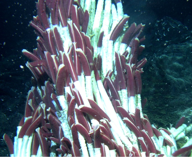
Riftia pachyptila, a polychaete typically found around hydrothermal vents have an organ with chemosynthetic bacteria instead of a gut.
(Photo credit: Wikimedia Foundation)

            So, what does this mean, what can be gained from this research? Well, Dick believes that this shows that these viruses are actually very important in the long term survivability of these bacteria. Dick and his team believe that these viruses act as “agents of evolution”, allowing for gene transfer between the chemosynthetic bacteria, a theory first proposed about photosynthetic bacteria found in the shallows by David Garrison, a program director for the National Science Foundation. Without these viruses, the bacteria would rarely receive gene transfer, and a disease could cause catastrophic damage to the bacterial population, which could cause an entire collapse of the hydrothermal vent community, since they rely on these bacteria for primary production.

References: 

Sunday, April 27, 2014

New Carnivorous Sponges

http://image.wistatutor.com/content/animal-kingdom/sponge-anatomy.jpeg
The above image shows the typical anatomy of a sponge.  These use the whip-like tails (flagella) of their collar cells to create a current of water.  This current of water allows the sponge to filter out bacteria and other food.  However, four new species of sponges (two species of Asbestopluma and two species of Cladorhiza) have adapted a different feeding strategy.  These new species inhabit the food-poor waters of the deep sea in the northeast Pacific.  Therefor it is not cost-effective for them to have this constant motion of cells to create a current.  Instead these sponges have a carnivorous feeding strategy.  When small crustaceans bump into the spiny sponge, they become trapped in their microscopic hook-like spicules.  A picture of the spicules from the species Abestopluma rickettsi can be seen below.

The sponge then proceeds to digest the crustacean.  A picture of the progressive decomposition of a crustacean by the species Asbestopluma monticola can be seen below.

These sponges are also often found in chemosynthetic habitats.  These are habitats where bacteria utilize the chemical-rich water as an energy source as opposed to sunlight.  The species Asbestopluma rickettsi was discovered in one of these habitats offshore of southern California where the bacteria use the methane that seeps from the seafloor.  Another species, Cladorhiza caillieti was discovered near a hyrothermal vent along the Juan de Fuca Ridge.  The fourth species, Cladorhiza evae was also discovered near a hydrothermal vent in the Gulf of California and is pictured below.

There is still much to learn about these sponges.  Researchers believe that these sponges may also be able to utilize the chemosynthetic bacteria as an additional energy source to the crustacean prey.  Only further research with these sponges will be able to confirm that hypothesis or not.


References:

  1. http://www.biologyjunction.com/sponges__cnidarian_notes_b1.htm
  2. http://biotaxa.org/Zootaxa/article/view/zootaxa.3786.2.1/7741
  3. http://www.eu-hermione.net/science/chemosynthetic-ecosystems


Better ways of protecting Coral Reefs?

With coral reefs being destroyed by bleaching and ocean acidification new methods of conservation must be approached.  One of these approaches is by not putting all efforts into the coral reefs directly buy by better conserving the land around the reef.  Researchers Carissa Klein, Stacy Jupiter, Matthew Watts and Hugh Possingham tested six different conservation techniques on coral reef ecosystems in Fiji.  The primary objective of each approach was to achieve terrestrial conservation or to minimize land-based runoff.  They also compared these results with other conservation plans on how well they benefit coral reef conditions.

The researchers found that when terrestrial conservation was the primary goal that the reefs benefited by  7.7-10.4% greater than other protected areas that weren't focused on terrestrial conservation.  However, 31-44% of the terrestrial conservation was not achieved which means that there could be a higher percentage of benefit if terrestrial conservation can be achieved better.  These results are being used by the Fiji's protected area committee to better define conservation boundaries on land.


Another method that is being researched is to take more of a gardening approach to the reefs.  Studies are being done on how successful coral reefs, grown in labs, that are modified to withstand current oceanic conditions.  Artificial reefs such as concrete structures are also being used.

A more abstract study looks into if electrical currents can stimulate coral reef growth.  On Vabbinfaru Island, Maldives a 12 meter metal cage that weighed 2 tonnes was placed in the ocean and a small electrical current ran through it.  The electric current stimulates a chemical reaction that draws calcium carbonate out of the water and gets deposited on the cage.  Studies have also been done that found that the electric current helps them filter and adapt to warmer conditions because less energy is needed to form a skeleton.  One of the steel cages used is hard to distinguish now because of all of the colorful reef growth on the cage.

Unfortunately using these steel cages on a global scale would be extremely costly.  The only benefit of these artificial reefs would be from tourist areas or areas that are hit by temporary damage such as oil spills or boat impacts.  Many of the coral reef researches believe the only way to save the reefs is to greatly slash carbon dioxide levels across the globe, but they don't see that being accomplished.  Researcher Peter Sale said "By 2050, we may still have corals, and things we'll actually call 'reefs', but they will be massive limestone structures that were built in the past, with tiny patches of living coral struggling to survive on them." He adds "The world will go on without reefs, but its going to be very much inferior to the planet we have now."

References:
http://www.sciencedaily.com/releases/2013/12/131217155329.htm
http://www.sciencedirect.com/science/article/pii/S0308597X13002261#
http://www.bbc.com/future/story/20120905-save-our-dying-coral-reefs
  • Saturday, April 26, 2014

    Barreleye Fish

    In our last class lecture, we discussed much about the adaptations of deep sea animals. This fascinated me because the mystery behind these creatures is worth looking more into. While scrolling through an article discussing the 25 most terrifying deep sea creatures, number 8 struck me as truly interesting. This organism is known as the barreleye fish. In the family Opisthoproctidae, it is scientifically known as Macropinna microstoma, and more commonly known as the spook fish. The reasoning behind its common name can clearly be seen below through the obvious observation that they have a transparent dome head, which indeed looks strange and spooky.  The barreleye fish have two upward facing eyes that allows them to specifically search for potential predators around them. What we would think would be eyes on this species (which are the two spots on its mouth) are actually nasal organs (equivalent to the nose in humans). I found that fact interesting because it definitely made me think that the two spots on its face were its eyes, when actually its eyes are the two green glands in the transparent portion of its head. The history of its existence has been described since 1939 but it wasn't until about 2008 that the true understanding of this species had been further discovered.

    Originally thought to have eyes that are fixed in place to only look up above, it was believed by marine biologists for quite some time that this was their disadvantage to not see what what in front of them. According to an article by Bruce Robison and Kim Reisenbichler, there is now a complete understanding that these fish eyes are tubular. Not only that, but the eyes are meant to peer through their transparent membrane to look straight above in search of potential predators. According to Robison and Reisenbichler, they used machinery known as remotely operated vehicles to study these deep sea fish off the coasts of central California. They reportedly caught one and examined its physiology. Their flat fins allow them to take advantage of being nearly motionless in the water and to maneuver efficiently. This is interesting because it allows them to save their energy. 

          Figure 1. The barreleye fish with its transparent head and tubular eyes (as green glands) shown above.

    In addition to their strange physiology of their eyes, the fact remains that their eyes show green pigments when the remotely operated vehicles (ROVs). These pigments are speculated to filter out all the sunlight above them and allow them to detect prey through bio-luminescence. This is a very interesting adaptation because it allows this species to stay protected by seeming quiet and vulnerable. Its small mouth makes it a predator who relies on getting energy/food effectively. The barreleye fish feed on jellyfish, which is effective and a positive thing considering they live within the same habitat as the jelly fish at these deep depths (2,000-2,600 feet). According to this article, Apolemia colonial jellyfish have stinging tentacles that can extend as long as thirty-three feet long, which can be deadly to other organisms within that area. As means to have a defense mechanism, the barreleye fish have adapted in protecting its eyes with a the ability to avoid the tentacles and also to rotate its overhead eyes. The fact that they can protect themselves in this way shows their ability to have adapted to the challenges of the deep ocean. The fact that it was the Monterey Bay Aquarium who discovered this species fully intact for the first time in its dome shaped head. I think what really was interesting to learning about this species was the fact that there are so many more organisms in the deep ocean that I hope to learn more in the future. 

    B.H. Robison and K. R. Reisenbichler. Macropinna microstoma and the paradox of its tubular eyes. Copeia. 2008, No. 4, December 18, 2008.

    Monterey Bay Aquarium Research Institute. Researchers solve mystery of dep-sea fish with tubular eyes and transparent head. February 23, 2009. 
    http://www.mbari.org/news/news_releases/2009/barreleye/barreleye.html 


    It's a Squid... It's a Worm.. It's a Squid Worm?

    Have you ever seen a creature so disgusting or bizarre that you were in shock? This is what happened to researchers at Woods Hole Oceanographic Institute. In a November 2010 expedition to the deep, marine scientists thought they had seen a worm eating a squid, or a squid eating a worm. This sight turned out to be neither of these things, but in fact a brand new species called the squid worm from the genus Teuthidodrilus. The squid worm is living proof that there are a massive array of undiscovered and incredibly strange organisms living in the largely unexplored depths of the ocean.

    http://news.nationalgeographic.com/news/2010/11/101124-squid-worm-new-species-science-teuthidodrilus-biology/

    The squid worm feeds on plankton, uses bristles to swim, and has pseudo-arms on its head which are primarily used for sensory function. The squid worm can grow to lengths of up to 9 cm. The squid worm resides in the depths of the ocean and has been found as deep as 6,200 meters in the Celebes Sea. Even the marine scientists responsible for the discovery were very surprised by the appearance of the squid worm.

    Perhaps the most perplexing thing about this organism is the fact that it is so common. This worm appears to be very abundant at the depths. This speaks volumes about our knowledge of the creatures that reside in deep marine ecosystems. With recent findings of creatures that are common and incredibly large creatures that would seem impossible to miss such as the giant squid, our knowledge of what exists on our own planet seems limited.

    The hadal zone of the ocean, named after Hades in Greek mythology, begins at about 6,000 meters - well within the range of the squid worm - and continues to the point where life no longer exists in the depths of the sediments. Currently, there is enough unexplored space in the hadal zone to equal roughly the size of Australia. This seems absurd considering our exploration of the moon and the heights of the earth such as Mount Everest.

    The ocean should not remain an enigma for very much longer. Advances in technology allows for scientists and unmanned vessels to explore the depths much more readily. James Cameron recently completed a voyage to the depths of the Marianas Trench, the deepest solo dive on record, and there is a current expedition exploring the Kermadec Trench which is one of the deepest and coldest trenches in the world. The outlook for future exploration is bright. The depths are predicted to contain diversity matching that of the coral reefs. One thing is certain, in the near future we will have a much better knowledge of the deep.

    References:

    Howard, Jacqueline. "Researchers Take On Kermadec Trench Dive To Find Unknown Deep-Sea Creatures." The Huffington Post. TheHuffingtonPost.com, 13 Apr. 2014. Web. 26 Apr. 2014. <http://www.huffingtonpost.com/2014/04/13/kermadec-trench-dive_n_5140725.html>.
    Sample, Ian. "'Squid Worm' Emerges from the Deep." The Guardian. Guardian News and Media, 24 Nov. 2010. Web. 26 Apr. 2014. <http://www.theguardian.com/science/2010/nov/24/squid-worm>.
    Than, Ker. "James Cameron Completes Record-Breaking Mariana Trench Dive." National Geographic. National Geographic Society, 09 Mar. 0040. Web. 26 Apr. 2014. <http://news.nationalgeographic.com/news/2012/03/120325-james-cameron-mariana-trench-challenger-deepest-returns-science-sub/>.
    "To Hades and Back: Exploring the Deepest Part of the Ocean | Expeditions, Scientific American Blog Network." Scientific American Global RSS. N.p., n.d. Web. 26 Apr. 2014. <http://blogs.scientificamerican.com/expeditions/2014/04/17/to-hades-and-back-exploring-the-deepest-part-of-the-ocean/>.

    Tuesday, April 22, 2014

    Sea Star Wasting Syndrome

    There is a new threat to sea stars called ‘sea star wasting syndrome’, which is responsible for mass killings of these important keystone species.  In November 2013, the disease killed up to 95 percent of the sea star populations from Alaska to Orange County.  Little is known about the origins of the syndrome, or even what causes it.  Scientists are trying to determine the cause of this lethal disease before time runs out.


    Typically, a sea star infected with the syndrome will have lesions that appear in the ectoderm followed by decay of tissue surrounding the lesions, which leads to eventual fragmentation of the body and death (see picture above).   A deflated appearance can precede other morphological signs of the disease.  “True” wasting disease will be present in individuals that are found in suitable habitat, often in the midst of other individuals that are affected.  The progression of wasting disease can be rapid, leading to death within a few days, and its effects can be devastating on sea star populations. 

    “They essentially melt in front of you,” said Pete Raimondi, chairman of the Department of Ecology and Evolutionary Biology at UC Santa Cruz's Long Marine Lab.  The University of California Santa Cruz is currently mapping all events along the West Coast, and people are encouraged to report these sea star wasting events to them.  They classify the syndrome into four categories, with 1 being mild, and 4 being severe.  Pictures and descriptions for the severity of the syndrome can be found here.   

    At first, the disease only infected one species, Pycnopodia helianthoides, also known as the sunflower star.  Then the disease began to affect a more common sea star species, Pisaster ochraceus (Robert Paine’s keystone species).  Now, there are about 12-15 species that are dying along the West Coast from sea star wasting syndrome.  And wild sea stars are not the only ones in danger-- in September 2013, sea stars in an aquarium at the Gulf of the Farallones National Marine Sanctuary visitor center at the San Francisco Presidio contracted the syndrome in water pumped from the ocean.  Eels, sculpin and anemones that were in the same aquarium were unaffected.

    The probable cause of the disease on the west coast of the US is caused by a pathological agent, such as bacterium (vibrio), although a recent wasting event on the east coast of the US has been attributed to a virus.  Sea star wasting events have also occurred from British Columbia down to the Gulf of California, the Mediterranean and the North Atlantic coast of North America, but not in the Southern Hemisphere.  Some researchers have suggested that Fukushima could be a cause, but the sea stars are being affected on the east coast, so this is not likely.  The ultimate cause is not clear although such events are often associated with warmer than typical water temperatures as was the case for the major die off in southern California in 1983-84 and again in 1997-98.  Sea stars are susceptible to bacterial infection, and warmer water boosts bacteria growth, Raimondi said.  

    If the cause for sea star wasting syndrome is not uncovered, ecosystem balance can be disrupted with the disappearance of sea stars.  As we have learned in class, removing Pisaster ochraceus from tide pools causes unchecked population growth of mussels and other organisms.  Also, with global climate change, temperatures will rise in the ocean, which could have significant or even profound effects on populations.  Hopefully the cause of sea star wasting syndrome can be solved before it is too late.

    Monday, April 14, 2014

    Elephant Seals and Swine Flu

    http://media-2.web.britannica.com/eb-media/96/99696-004-058EB9C8.jpg
    This awkward looking fella' belongs to the species Mirounga angustirostris, or more commonly, the northern elephant seal.  Male elephant seals can grow up to 14 feet in length, and weigh over 5,000 lbs!  They inhabit the west coast of North America and can travel as far as Japan.  They are excellent swimmers for this is how they get most of their food; hunting fish and squid.  At extremes, elephant seals can dive to depths of 1,500 meters, and stay under water for a whopping two hours!  They can also travel over 12400 miles, annually. Though these giant marine mammals may seem tough, they are susceptible to many diseases.

    In 2010, it was discovered that 28 elephant seals had traces of H1N1 anitbodies, suggesting that they were or had been infected with the virus while 2 were tested positive for the infection itself.  H1N1, you may better remember as swine flu, originated from pigs, but had emerged in humans in 2009 as a huge pandemic.  Elephant seals were tested at the beginning of 2010, and tested negative, but upon return from the sea in spring, tested positive.  The figure below maps out this phenomena.
    Evidence of exposure to H1N1 in northern elephant seals off central California in 2010 to 2011
    In the same study, two adult female elephant seals that tested positive for the virus had their paths tracked in 2008 and 2010.  The data collected can be seen in the figure below.
    Tracking data from two adult female elephant seals that tested positive for H1N1 from 2008 and 2010
      This data shows that both seals took similar paths out to sea in 2008 and 2010.  Infection was detected in both seals within days of their return from sea suggesting that they were exposed before their return.  The question is; Where exactly were they and exposed and how?  Though this is not the first time that marine mammals have tested positive for strains of influenza, that question still remains unclear.


    References

    1. Tracey Goldstein, Ignacio Mena, Simon J. Anthony, Rafael Medina, Patrick W. Robinson, Denise J. Greig, Daniel P. Costa, W. Ian Lipkin, Adolfo Garcia-Sastre, Walter M. Boyce. Pandemic H1N1 Influenza Isolated from Free-Ranging Northern Elephant Seals in 2010 off the Central California Coast. PLoS ONE, 2013; 8 (5): e62259 DOI: 10.1371/journal.pone.0062259
    2. Goldstein T, Mena I, Anthony SJ, Medina R, Robinson PW, et al. (2013) Pandemic H1N1 Influenza Isolated from Free-Ranging Northern Elephant Seals in 2010 off the Central California Coast. PLoS ONE 8(5): e62259. doi:10.1371/journal.pone.0062259
    3. http://www.mnh.si.edu/mna/image_info.cfm?species_id=184
    4. http://www.sciencedaily.com/releases/2014/04/140408213619.htm